by Rachel Bissonnette, Graduate Book Conservation Intern, Weissman Preservation Center
The illustrated sequence in the 聚星圖 (Chʻwisŏng-to, A Narrative Figure Paintings about the Virtuous Gathering of and Xun Family) (c. 1677) album depicts the meeting of two late Han dynasty families, the Chen and Xun. There are three paintings in this manuscript attributed to the celebrated Korean painter, Jo Se-geol. Three other known versions of this illustrated sequence exist, but scholars speculate that this is the earliest version and was a working copy. Curators from the National Chuncheon Museum in South Korea contacted the Weissman Preservation Center to request technical analysis be performed on the Harvard-Yenching Library manuscript so that they could better understand the creation of the paintings (Fig. 1).
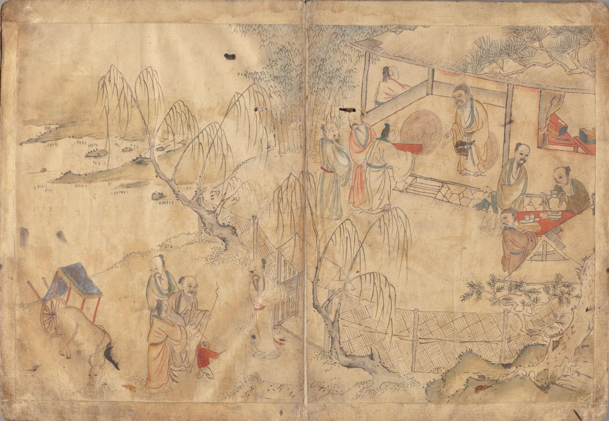
Figure 1. Second painting in the Chʻwisŏng-to illustrated sequence.
The team performing the analysis included: Debora Mayer, Helen H. Glaser Conservator; Katherine Beaty, Book Conservator; Kelli Piotrowski, Special Collections Conservator; and Rachel Bissonnette, Graduate Book Conservation Intern. The researchers used close observation, x-ray fluorescence (XRF) spectroscopy, and multispectral examination with the Video Spectral Comparator (VSC) to address the curators’ questions (Fig. 2). The curators were primarily interested in identifying the original composition of the painting because it appears the paintings may have been reworked or overpainted.
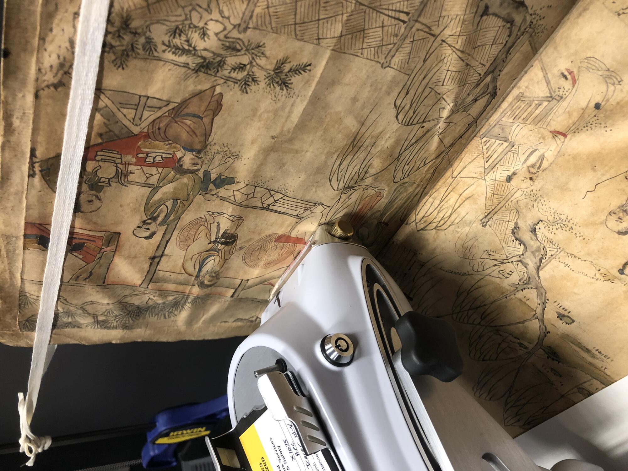
Figure 2. Using a Bruker TRACeR III-V XRF portable analyzer to examine the colorants.
Analysis focused on identifying four key colorants used to create the paintings: reds, greens, blues, and dark grays.
The primary red observed across the three illustrations is a bright red-orange, opaque pigment applied to the robes, tables, and cart rails (Fig. 3). XRF identified strong mercury peaks suggesting the presence of vermillion (mercuric sulfide) (Fig. 4).
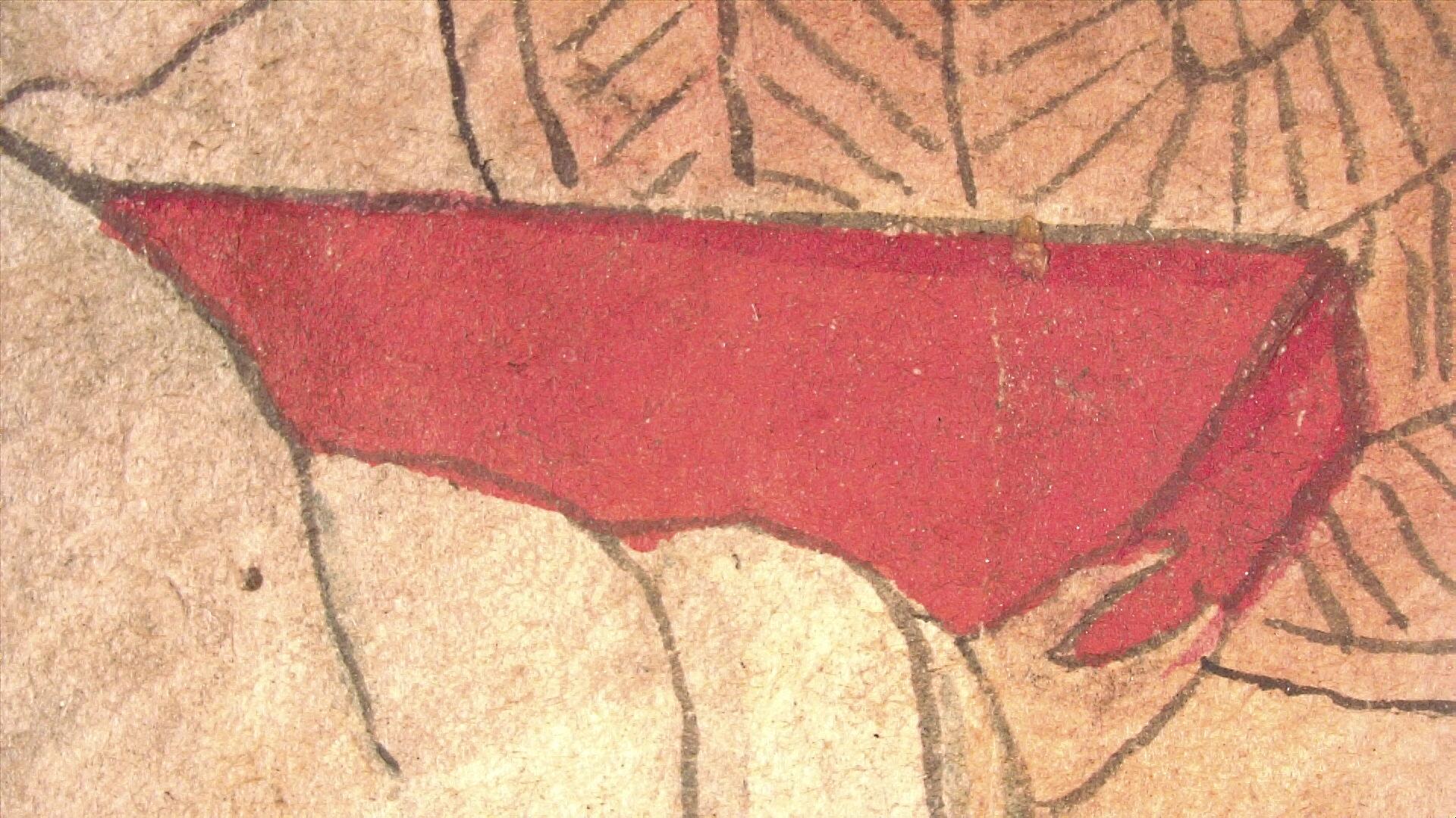
Figure 3. 5x magnification photomicrograph of a red tray in the second painting.
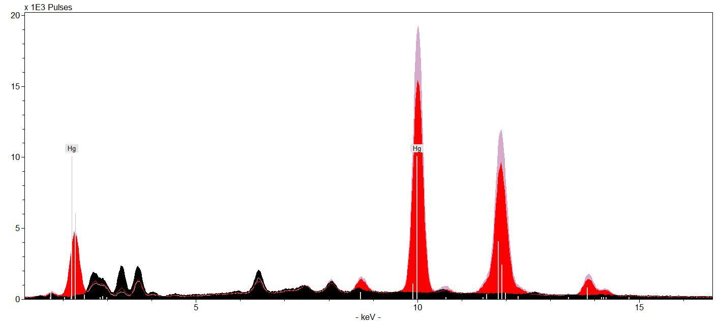
Figure 4. XRF spectra of red tables showing strong mercury signals.
Most of the green in the illustrations is present in the hills and vegetation applied as a wash (Fig. 5). XRF identified copper in the green hills, suggesting the presence of a copper containing pigment such as malachite (basic copper carbonate) or verdigris (basic copper acetate) (Fig. 6).
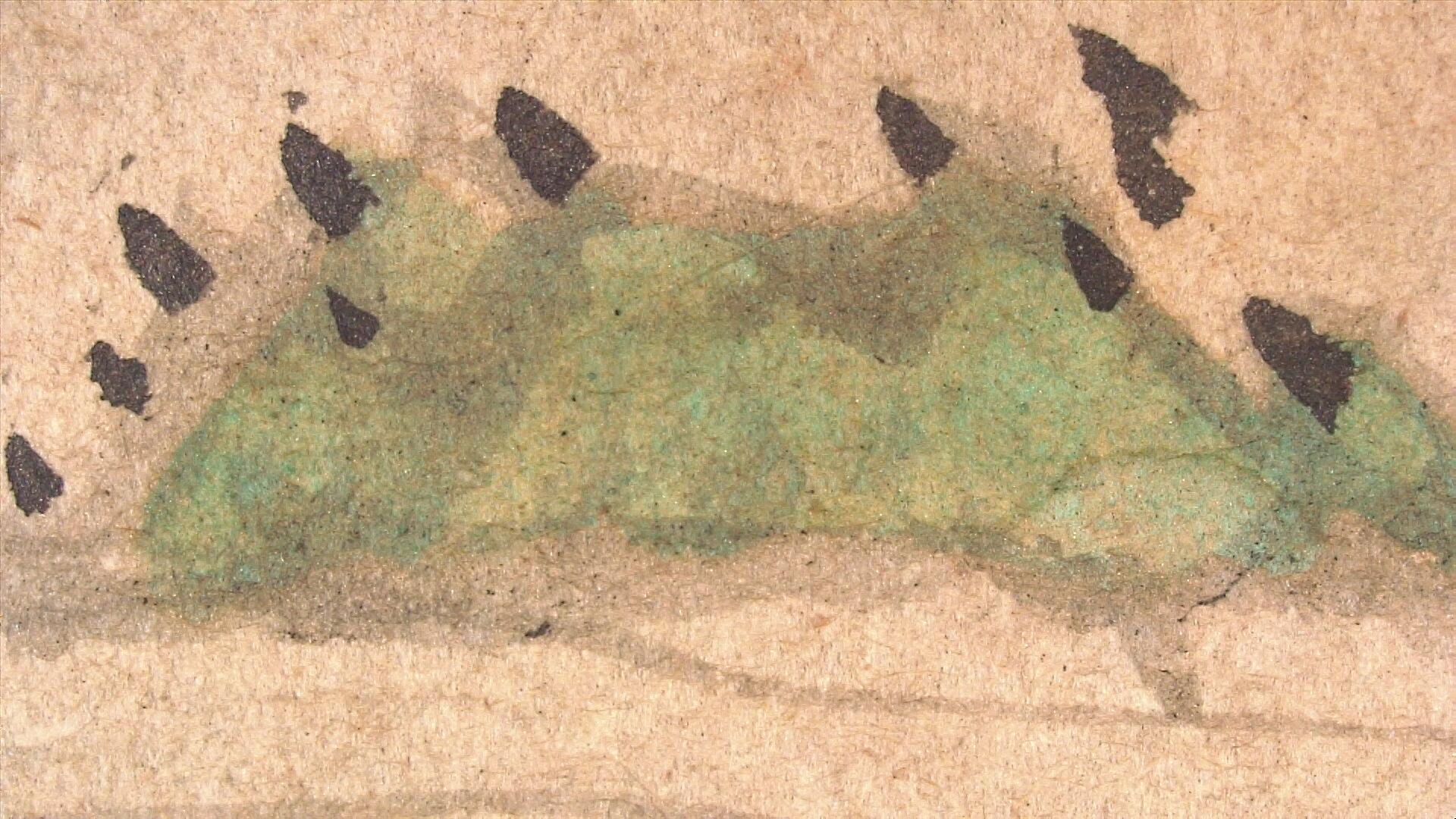
Figure 5. 6x magnification photomicrograph of pale green used in the hills.
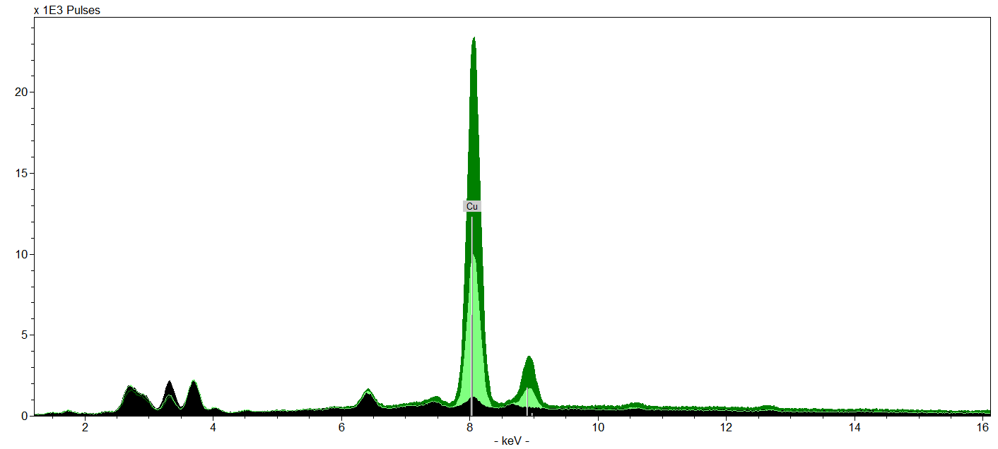
Figure 6. XRF spectra of green hills showing strong copper peaks.
Two distinct blues were observed in the illustrated sequence. A bright, thickly applied coarse blue pigment was used for the topknots of the figures, robe collars, and jackets (fig. 7). A different, more transparent blue-green was identified in the robes, sashes, and conifer needles (fig. 8). Spectra from XRF of the topknot, blue collar, and blue jacket had strong copper signals suggesting the presence of azurite (basic copper carbonate) (Fig. 9). XRF could not detect any strong signals from the blue-green colorant, implying organic media.
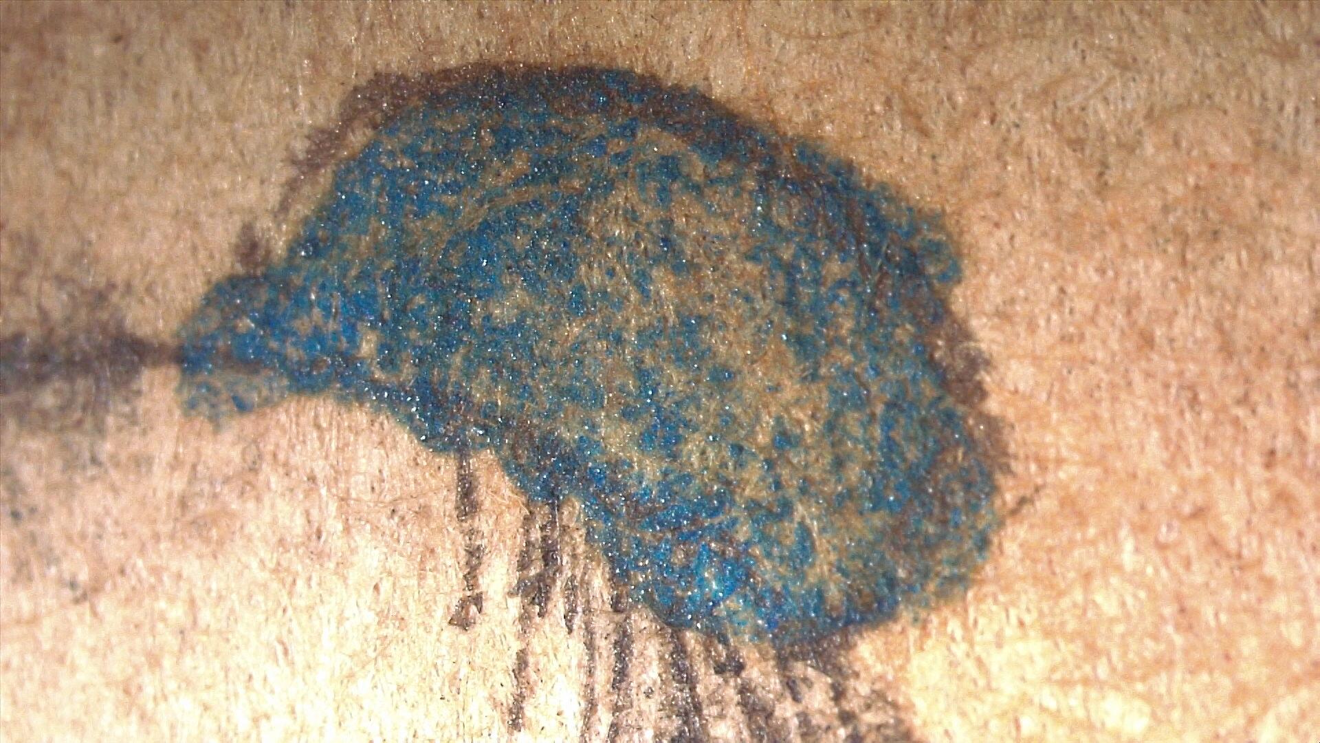
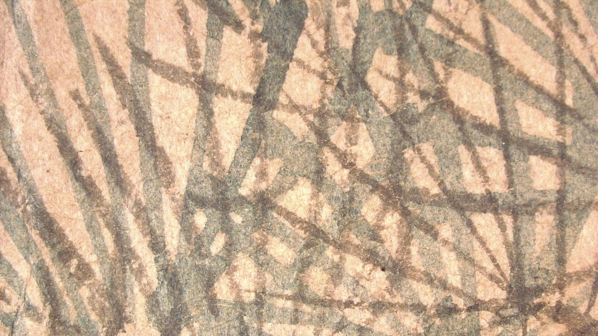
Figure 7-8. 17x magnification photomicrograph of bright blue colorant present in one of the topknots (left). 6x magnification photomicrograph of blue-green colorant present in the conifer needles (right).
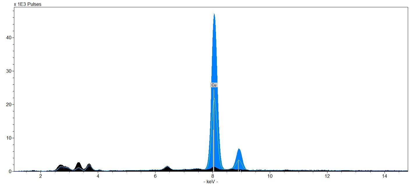
Figure 9. XRF spectra of child’s blue jacket shown in dark blue and blue topknot shown in light blue. Strong copper signals were detected in both spectra.
Multispectral imaging from the VSC corroborated the presence azurite in bright blue areas by comparison with reference spectra. This same technique was used to identify indigo in the robes and conifer needles (Figs. 10-11). Indigo is an organic blue dye sourced from the leaves of the plant species indigofera tinctoria.
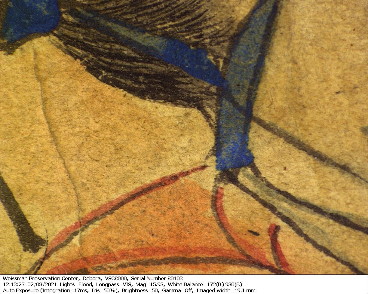
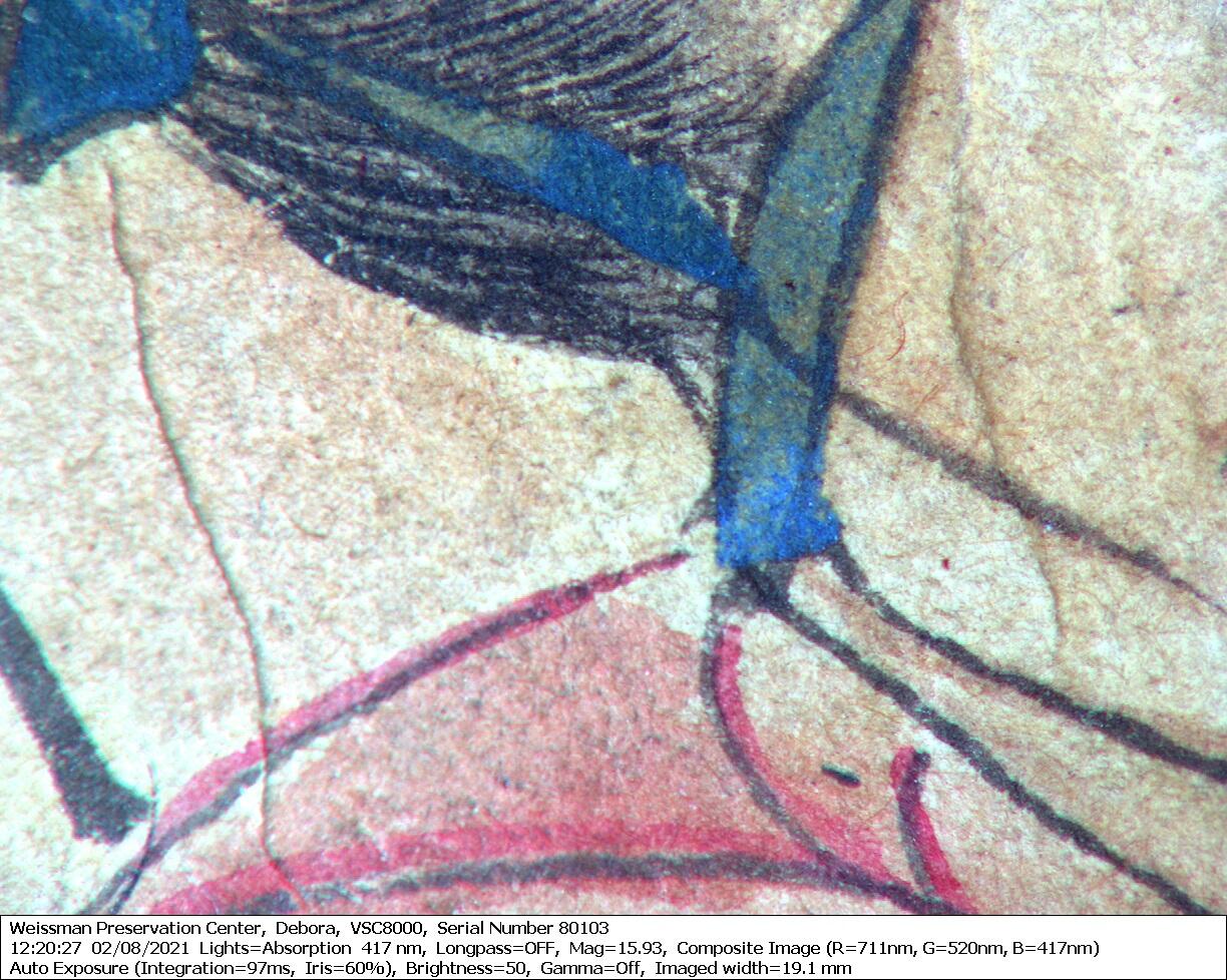
Figures 10-11. These two images captured at 16x magnification with the VSC show the difference in reflectance of the two blue pigments. In the visible light image (left) there is the bright blue colorant present in the topknot and collar of the robe, and a lighter blue wash in folds of the robe. In the false color image of the same area (right) blue observed in visible light used for the folds of the robe now appears to be pinkish-brown while the blue in the topknot and collar still appears blue. This suggests the two blues were created using different colorants.
The grays were the most challenging to analyze and remain mysterious. A grainy, dark gray colorant was used on the cart roofs and to accentuate the tea sets and book edges (fig. 12). A similar dark gray was observed in the facial features of some of the figures (fig. 13). The broad, dark gray marks in some of the faces are easily distinguished from the matte, finely applied black ink lines, which suggests the dark gray may have been a later intervention.
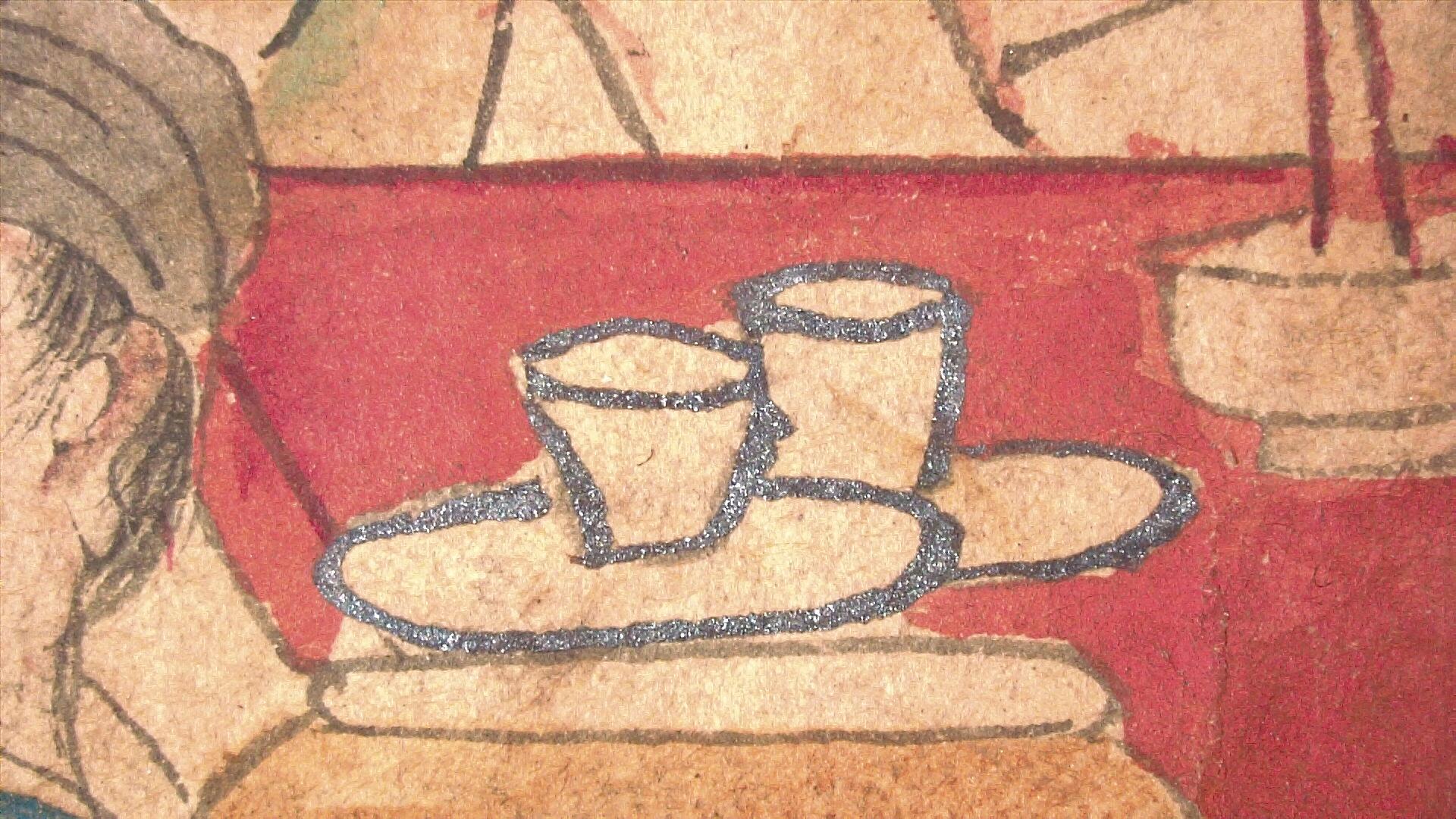
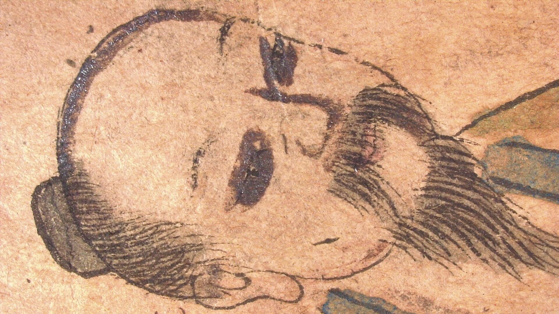
Figure 12-13. 5x magnification photomicrograph of the shiny gray colorant used on the tea set in second painting (left). 5x magnification photomicrograph illustrating the different ink and colorant applications observed in the faces (right).
Multispectral imaging from the VSC shows two distinct applications of media (figs. 14). The likely carbon-based black ink lines and the shiny gray. The only strong elemental peak discovered with XRF was lead (fig. 15). This suggests the dark gray is discolored lead white or red, however this does not preclude the presence of organic colorants that aren’t detectable with XRF.
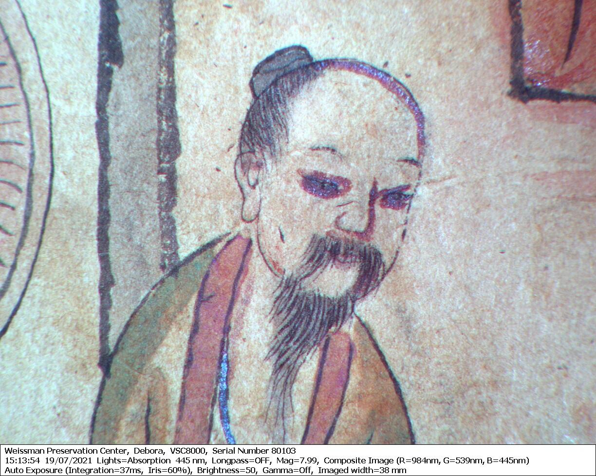
Figure 14. This false color image captured at 8x magnification with the VSC shows that the black and dark gray lines are not the same pigment composition by the differing color response— the black lines remain black while the dark gray lines on the top of the head and robe take on a blue-purple cast.
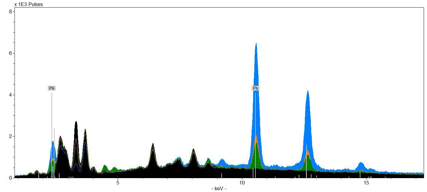
Figure 15. The green and orange regions represent spectra of faces with suspected dark gray overpainting. The blue spectrum is of the man in cart with white facial hair that has no evidence of the dark gray overpainting. All these spectra have strong signals for lead, possibly indicating a wash of lead white which may have discolored to dark gray.
The researchers could not conclusively identify the composition of the dark gray, nor could they definitively say if it was part of the original composition. The dark gray, which may be a discolored form of lead, could have been a later intervention, but the media analyzed is contemporary with the production date of the manuscript. All we can say for certain is lead is a component of the dark gray and it is not the same carbon-based black ink that was used for the lining drawing—as observed with the VSC.
It was a pleasure to work so closely with this manuscript and use technical analysis to unravel some of its mysteries.


